

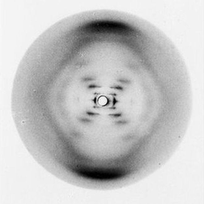
“The moment that launched the bio-molecular age: Celebrating the anniversary of history’s most consequential X-ray photograph.”
May 2nd to the 6th next year will mark the 75th anniversary of the X-ray diffraction image of DNA known as Photo 51. It would be nearly a year before Maurice Wilkins would infamously show the image to Jim Watson, and shortly after, Watson and Crick - combining information from it and other sources - would successfully predict the double helical structure of DNA. The 75th anniversary of the discovery of the structure of DNA will be on April 25th 2028, both will be noted, perhaps the more important would be that of Photo 51, which is so widely considered today as presenting the singular evidence of the molecules double helical structure for the first time, while of course, the discovery by Watson and Crick and publication of it’s double helical structure is the more historically significant.
Photo 51 was taken by Ray Gosling, then a young doctoral student at King’s College’s Biophysics Laboratory, a special unit created by the Medical Research Council under the leadership of Sir John Randall in 1946. Though not a Nobel Laureate like his counterpart, Sir Lawrence Bragg at the Cavendish Laboratories, Cambridge, Randall was a distinguished physicist who had also made significant contributions to the war effort through the development of the cavity magnetron, which was fundamental to radar development — one of the most significant British scientific contributions of the war. It was Randall’s intention to develop the King’s unit as the world’s premier center for the study of the structure of important biological molecules using X-ray crystalographic methods, which had not previously been applied to this purpose. To lead the team, Randall hand-picked Maurice Wilkins to lead the work on DNA. Wilkins had been one of the select group of British physicists sent to work on the Manhattan Project, developing the world’s first nuclear weapon. While Cambridge was focused on the structure of hemoglobin, led by Max Perutz and John Kendrew, Wilkins had chosen the equally complex structure of deoxyribonucleic acid, a molecule widely regarded as the fundamental basis of life. During the early years of setting up the lab, Wilkins designed and built the specialized X-ray diffraction cameras needed to photograph biological molecules and successfully secured the only then-available source of crystalline DNA pure enough to yield useful X-ray diffraction results.
Wilkins had trained his doctoral student assistant, Raymond Gosling, to use his camera and had prepared the fiber from the crystalline DNA so that images could be taken. What happened next is best described as a very British Affair, paralleled as a story of mythmaking, perhaps only by John Ford’s classic western “The Man Who Shot Liberty Valance.” The myth surrounding Photo 51 and the specific role it played in the discovery of the double-helical molecular structure of DNA is covered in the next Ariadne of this series, “The Betrayed Legacy of Rosalind Franklin.” Here, it’s sufficient to say that Wilkins left the lab for a short vacation, and upon his return, he learned that Sir John had hired a highly skilled crystalographer, Rosalind Franklin, and assigned Ray Gosling and the DNA exclusively to her.
What is Photo 51?
It could easily pass as a minimalist abstract painting, but it is, in fact, the first convincing scientific evidence of the DNA molecule’s double-helical structure. As a result, the original, a 600-millimeter-square X-ray image sandwiched between glass plates for projection, holds immense significance in scientific history and is a foundational image of molecular biology. Its title a simple reference to its position in a sequence of X-ray diffraction studies being undertaken at the laboratory. Its significance, when compared with other DNA images taken at the time, lies in being the first X-ray diffraction image of the B form of DNA, showing the distinctive X-ray pattern now accepted as uniquely indicative of the molecule's helical structure.
The terms A and B refer to the forms of DNA fibers used in X-ray diffraction, which produced images that varied with relative humidity at the time of exposure; exposures sometimes lasted several hours. The A-form, produced under low humidity, is a shorter and wider double helix whose complex diffraction pattern obscured the helical signature, while the B-form, produced under high humidity conditions, gave the unmistakable "X"-shaped diffraction pattern — famously captured in Photo 51 by Franklin and Gosling — that directly revealed the helical backbone geometry Wilkins had long suspected.
Photo 51 was not taken by Rosalind Franklin, but by Raymond Gosling. It is very important to note this distinction because, at the time, she neither recognized nor accepted the double-helical interpretation of the molecule’s structure proposed by Maurice Wilkins. Since the photo was not taken by Franklin, it was not her property and was, in fact, widely shared with others of the King’s team by Gosling, who had obtained Franklin’s consent to do so.
Before Franklin’s arrival at the lab, Gosling had been Maurice Wilkin’s student and had been trained by him to use the camera Wilkins had designed and constructed for the X-ray diffraction studies. He had shared with Franklin’s knowledge and consent with Wilkins and other members of the King’s team.
Gosling was first a doctoral student of Wilkins, whom he had been assigned to work with, and was assigned to work with Rosalind Franklin when she joined the DNA research group at King’s College, University of London. Franklin also took over the supervision of Gosling's doctoral thesis.
At the time, the King's laboratory was far in advance of any other in the world in the x-ray diffraction study of DNA. Additionally, the DNA samples available for making the diffraction images in the laboratory were limited to two, one of which was of superior quality. Franklin’s work was crucial to distinguishing the differences between the A and B forms, though she did not subscribe to the view of Wilkins that DNA in both forms was helical in structure.
The photo is considered iconic and a significant moment in the history of molecular biology for both the experimental process used to obtain it, as well as its legendary association with the discovery of the double helical structure of the molecule made famous by James Watson's personal account of the discovery, "The Double Helix."
Photo 51: From Calculation to Confirmation
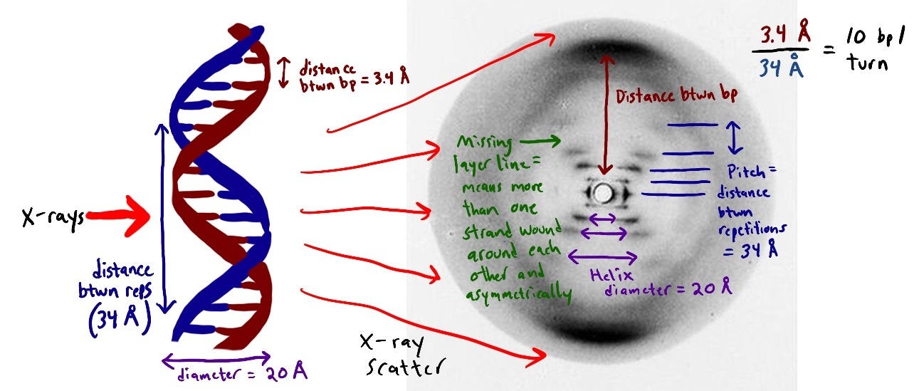
Creating Photo 51 was no ordinary photography. A tiny sample of hydrated DNA was mounted, and an X-ray beam was directed at it for 62 hours. The resulting striped cross shape, visible in the image, held the key to DNA’s structure. The distinctive striped cross shape in the picture held the critical information about DNA’s helical nature and is clearly presented in the illustration produced by Emily Willoughby (above).
When Maurice Wilkins showed this image to Watson, who was visiting him at King’s, Watson's reaction was profound: "My mouth fell open, and my pulse began to race' he wrote in The Double Helix. The revelation of the double helix structure was a pivotal moment in science. The image provided the final clue that enabled scientists Maurice Wilkins, James Watson, and Francis Crick to piece together disparate lines of research and understand that DNA exists as a double helix. It was this revelation that fundamentally changed our understanding of genetics and laid the groundwork for modern molecular biology.
The iconic X-ray diffraction image known as Photo 51 was captured by Raymond Gosling between May 2 and May 6, 1952. Using a micro-focus X-ray tube and a hydrogen-filled camera to maintain 75% humidity, Gosling exposed a single fiber of calf thymus DNA for approximately 62 hours. This technical achievement was the empirical confirmation of a structural theory already in development at King’s College. Prior to the exposure, Alec Stokes, at the request of Maurice Wilkins, had utilized Fourier transform mathematics to calculate exactly what an X-ray diffraction pattern would look like if DNA were a helical molecule. Stokes’ calculations predicted that a helix would produce a characteristic “X” or cross-shaped pattern.
While Rosalind Franklin remained focused on the more data-rich “A” form for her mathematical derivations, viewing it as the more scientifically rigorous path, Gosling’s exposure of the hydrated “B” form provided the exact visual evidence Stokes and Wilkins had anticipated. As Franklin prepared to move to Birkbeck following her resignation from King’s, she was packing her materials and winding down her DNA research. During this transition, she consented to Gosling’s request to share the B-form data with the rest of the King’s biophysics team—including Wilkins, Stokes, and Herbert Wilson. Because the mathematical “X” had already been predicted by Stokes, the clarity of the B-form image produced by Gosling acted as the definitive proof that the double-helix model was the correct physical reality.
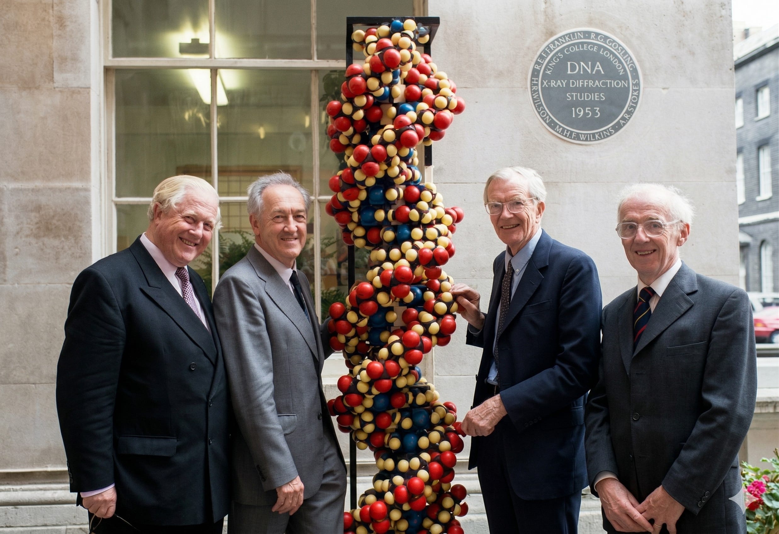
Why did Wilkins show Photo 51 to James Watson?
Gosling's image of the B form was taken in May 1952, a full year ahead of Watson's visit to King's, during which, in his recounting, he met with both Franklin and Wilkins. Almost a year went by before it was shown to Watson and Franklin, and she was leaving for Birkbeck College, leaving her DNA work behind. She had long given Gosling permission to show Photo 51 to others in the lab, particularly Wilkins, Alec Stokes, and Herbert Wilson. The diffraction images were shared within the group, not least because interpreting the molecular structure required collaborative calculations. Within the group, Wilkins was heavily in favor of a helical structure. Franklin could not be convinced of this, and the differences between the A and B forms left enough room for her doubt.
Would Watson and Crick have been able to solve the structure of DNA without Watson having seen Photo 51?
Crick has been reported to have claimed that the information from photo 51 was not as central to the discovery of the double-helical structure of DNA as suggested, at least by Watson's account in his book The Double Helix. Wilkins' account of his meeting with Watson offers a contrasting view of Watson's reaction to Franklin's resistance to accepting a helical structure for DNA, a view corroborated by what Franklin herself told Aaron Klug years later. By this time, both Wilkins and Watson and Crick knew that helical phosphate chains were on the outside of the bases, and because of Chagaff's work, Linus Pauling, together with Robert Corey, would likely have made another attempt at the structure as soon as they had additional data.
Would Franklin have solved the structure by herself?
Francis Crick is reported to have said that if he and Watson had not successfully modeled the structure of DNA in April 1953 (which had been completely derived from the experimental work of others), the double-helical structure would not likely have been proposed by others for another decade. This view perhaps reflects more on the opinion that Watson and Crick held for their competitors than the documented evidence. Rosalind Franklin decided to leave her DNA work behind when she sought to transfer to join J.D. Bernal's group at Birkbeck College, focusing on viral structure. She had recently moved when Watson and Crick assembled their second model, which Franklin traveled with the team to view. Sir John Randall, Maurice Wilkins, and Franklin were accompanied by Ray Gosling and Alec Stokes. After it was agreed that Crick and Watson would publish their proposed structure in Nature, it was also agreed that Wilkins and Franklin would submit supporting papers based on the work at King's. Franklin had consented to supervise Gosling’s doctoral thesis despite her moving to Birkbeck; aside from that, her research interests were now elsewhere. There’s no evidence that she held a desire to go back to work on DNA, so it's difficult to speculate how she would get her attention back. Mostly, it's been suggested by Aaron Klug, her colleague at Birkbeck and life advocate, that Franklin's various notes could be interpreted as indicating she was close to solving the molecule's structure. In an interview, Klug quotes Frankiin as having said to him on more than one occasion that she could "kick myself for not having given in to a helical structure sooner' for both the A and B forms.
Franklin went on to complete work on the structure of viruses at Birkbeck, where she worked until her death. She never raised concerns over the use of the data derived from her presentation at King’s attended by Watson, or that he had seen the photo 51 with the distinct X pattern of the B form of DNA.
She was an extremely intelligent woman, and it's unlikely she did not know, either through Gosling or from the discussion about viewing the model in the Cavendish Laboratory, that data from her work had been incorporated into it. This data was valuable but not absolutely essential for the proposed Watson-Crick double-helical structure.
However, Linus Pauling and Robert Corey had proposed a triple-helical structure for DNA, comparable to Watson and Crick's first attempt, without the benefit of any diffraction images from King’s. Pauling had unsuccessfully written to Wilkins, asking to see the X-ray diffraction studies. Once Pauling’s travel restrictions had been lifted by the State Department, and he would have been free to visit King’s or for King to share their data, it's very likely that Pauling and Corey would have succeeded in constructing a double-helical model.
If Watson and Crick had not obtained the information they derived from Watson’s viewing of photo 51 and her colloquium presentation and research report, would Franklin have solved the structure by herself?
Francis Crick is reported to have said that if he and Watson had not successfully modeled the structure of DNA in April 1953 (which had been completely derived from the experimental work of others), the double-helical structure would not likely have been proposed by others for another decade. This view perhaps reflects more on the opinion that Watson and Crick held for their competitors than the documented evidence. The evidence suggests that Linus Pauling, having made one unsuccessful attempt, built on the poor X-ray diffraction pictures he was able to get from Astbury, would certainly have closed in on the base pairs in the middle and sugar-phosphate chains on the outside approach sooner rather than later, and certainly within months rather than years.
Franklin had recently moved to Birkbeck when Watson and Crick assembled their second model, which Franklin traveled with the King's team to view. Sir John Randall, Maurice Wilkins, and Franklin were accompanied by Ray Gosling and Alec Stokes. After it was agreed that Crick and Watson would publish their proposed structure in Nature, it was also agreed that Wilkins and Franklin would submit supporting papers based on the work at King’s. Franklin had consented to continue supervising Gosling’s doctoral thesis despite her move to Birkbeck; aside from that, her research interests were elsewhere. There's no evidence that she wanted to return to work on DNA, so it's difficult to speculate how she would regain her focus. Mostly, it's been suggested by Aaron Klug, her colleague at Birkbeck and an advocate, that Franklin's various notes could be interpreted as indicating she was close to solving the molecule's structure. In an interview, Klug quotes Franklin as having said to him on more than one occasion that she could "kick myself for not having given in to a helicai structure sooner, for both the A and B forms. She never raised concerns over the use of the data derived from her presentation at King’s attended by Watson, or that he had seen Photo 51 with the distinct X pattern of the B form of DNA. She was an extremely intelligent woman, and it's unlikely that she did not know, either through Gosling or arising as a result of the discussion over viewing the model in the Cavendish Laboratory, that data from her work had been incorporated into the model. This data was valuable, but only one of several essential pieces of information needed to describe the proposed Watson-Crick double-helical structure.
However, Linus Pauling and Robert Corey had proposed a triple-helical structure for DNA, comparable to Watson and Crick's first attempt, without the benefit of any diffraction images from King’s. Pauling had previously unsuccessfully written to Wilkins, asking to see the X-ray diffraction studies. Once Pauling’s travel restrictions had been lifted by the State Department, and he would have been free to visit King’s or for King to share their data, it's very likely that Pauling and Corey would have succeeded in constructing a double-helical model.
What makes Photo 51 an iconic image?
Beyond its purely scientific impact, photo 51 has come to represent a portrayal of Rosalind Franklin as a mistreated Jewish woman scientist at a time when such discrimination was sadly commonplace. Her contributions towards the discovery of DNA have been argued to have been underappreciated, even maliciously ignored at the expense of her male colleagues. Specifically, James Watson’s writing of his personal account of the discovery of DNA, some ten years after her death, provided a misogynistic and denigrating portrayal of Franklin, with only Wilkins and Crick to defend her. Following the highly successful publication of his book "The Double Helix," Anne Sayre, a close friend of Franklin, particularly towards the end of her life, researched a book whose purpose was to counter the picture of her that Watson's account gave.
Published in 1975, Rosalind Franklin and DNA launched Franklin as a cause celebre for feminist scientists. Indeed, there was a wrong to be righted, but she was, I’m sorry to say, amongst a pantheon of important women scientists who had been overlooked and underacknowledged, particularly in the first half of the twentieth century. ln her book Sayre misrepresented Franklin’s difficulties at King’s created a false caricature of Wilkins as a thief of Franklins data and creating a myth that exploded into women scientist movement vilifying Wilkin's and elevating Franklin as a feminist icon robbed of fair accreditation for her scientific work to the point of un-personing Wilkins from the discovery for which he shared the Nobel Prize with Watson and Crick. At this moment, the discovery is commonly attributed to Watson and Crick, using Franklin’s data illicitly obtained through Wilkins, who did not have Franklin's consent to share it with Watson.
Though Crick and, much later, Wilkins published their own accounts of the discovery and the necessary historical research that countered the false narrative in Sayre’s book, that narrative persists and remains deeply embedded, much like the myth of John Ford’s movie western The Man Who Shot Liberty Valance. When Maurice Wilkins showed Photo 51 to “Honest Jim,” as he liked to be called, Watson's reaction as described in The Double Helix was profound: ‘My mouth fell open, and my pulse began to race.’ This image provided the missing link, allowing them to publish the groundbreaking paper on DNA’s structure in 1953. Crick, Watson, and Wilkins later shared the Nobel Prize. Watson's account of the taking of Photo 51 is not history. It is, by far, the better story.
© 2026 Farooq Hussain
Postscript
The discovery of the structure of DNA has been extensively written about, including many distortions that have neither helped to right the historical account nor to remedy the wrongs. Here, Ariadne looks only at how Photo 51 was taken to consider whether any X-ray diffraction of DNA at King’s could reasonably have been considered the intellectual property of an individual. Next, we consider how, in the attempt to right a wrong, Franklin’s true legacy has largely been sacrificed in response to a misogynistic and false portrayal of her as a person and a scientist in a book that has found its place among the best of twentieth-century American literature.
All history has relics associated with it. In this case, what now seems primitive equipment designed in-house and hand-built, including war-surplus components such as the X-ray source generator tubes, is bafflingly crude in construction, and we are lucky that, as molecular biology aggressively progressed through the second half of the twentieth century, some of these artifacts have been saved and are in museums and archives. The equipment used to create Photo 51 is described in the references presented in the Notes below.
Notes
The articles and videos below are well-researched accounts of how Photo 51 was made:
Browell, Geoffrey. "DNA at King's: The Making of Photograph 51." YouTube, uploaded by King's College London, 1 Nov. 2021, www.youtube.com/watch?v=F041-7Bmheo.
Sutton, Brian. "The Story Behind Photograph 51." King's College London, 14 Apr. 2023, www.kcl.ac.uk/the-story-behind-photograph-51.
The Making of Photo 51 https://www.kcl.ac.uk/discovering-the-structure-of-dna Molecular Biology Published: 26 April 1974
The Making of Photo 51, YouTube